
Leica Laser Microdissection 7
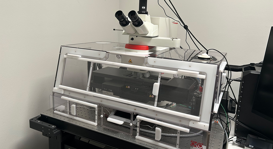
Microdissection allows one to select and isolate part of a sample and use it for further studies(RT-PCR, protein analysis, selection of live cells). Laser microdissection can also be used withlive samples to study recovery after DNA damage and protein recruitment after photodamage.
Specifications
|
Imaging |
Widefield |
|
Camera |
Leica K7 |
|
Stand |
LMD7 basic stand |
|
Stage |
Motorized in XYZ |
|
Software |
LAS X Premium |
|
Add. features |
Incubation system |
Objectives
|
Objective |
Mag./ NA |
Contrast |
WD |
Product No |
Use |
|
Plan 4X/0.10NA |
4x / 0.1 |
TL-BF, FLUO |
26.2 |
13613240 |
collector observation |
|
UVI 5x/0.12 Microdissection |
5X/0.12 |
TL-BF, FLUO |
13.5 |
11518151 |
laser microdissection |
|
HC PLFLUOTAR10x/0.32 PH1 |
10x /0.32 |
TL-BF, TL-DIC,TL-P, TL-PH,FLUO |
11.2 |
11506537 |
laser microdissection |
|
HC PL FL L20x/0.40 CORRPH1 |
20x / 0.4 |
TL-BF, TL-DIC,TL-P, TL-PH,TL-IMC, FLUO |
7.5-6.2 |
11506243 |
laser microdissection |
|
HC PL APO20x/0.80 |
20x / 0.8 |
TL-BF, TL-DIC,FLUO |
0.4 |
11506529 |
imaging |
|
HC PLFLUOTAR L40x/0.60 CORRXT |
40x / 0.6 |
TL-BF, TL-DIC,TL-P, TL-IMC,FLUO |
3.3-1.9 |
11506208 |
laser microdissection |
|
HC PLFLUOTAR L63x/0.70 CORRXT |
63x / 0.7 |
TL-BF, TL-DIC,TL-P, TL-IMC,FLUO |
2.6-1.8 |
11506222 |
laser microdissection |
Light sources
- For Brightfield TL: LED (PMax = 15 W) amp housing
- For Fluorescence
- For Dissection: Laser Diode-Pumped Solid-State (DPSS),
- 349 nm (ultraviolet, UV),
- 120 μJ average energy pulse,
- repetition rate 10-5000 Hz
- Laser aperture control continuously adjustable
- UV offset freely adjustable
Filter Cubes
|
Fluorochrome |
Filter No |
|
LMD-DAPI |
11505261 |
|
LMD-GFP |
11532651 |
|
LMD-Alexa 594 |
11505262 |
|
LMD-Cy5 |
11505269 |
|
LMD-BGR |
11513911 |
Collection device fittings
- 4 slides specimen holder
- multi-collector for up to four 0.2 mL or 0.5 mL standard PCR tubes with flat cap
- one 1.5 mL PCR tube with filled cap for direct contact pick-up
- one 8-strip tube cap
- full-skirted and semi-skirted 96-well PCR plates
Dissection Modes
- Draw & Cut
- Move & Cut (direct online cutting)
- PtoP (point-to-point)
- Draw & Scan (dot dissection scan)
- LMD screw (cut subsequently in Z-direction)
- Shadow Cuts (parallel draw and dissection)
Applications
- Microdissection of cells and tissue areas, aided by machine learning software
- Multicolor fluorescence imaging
- Transmission imaging