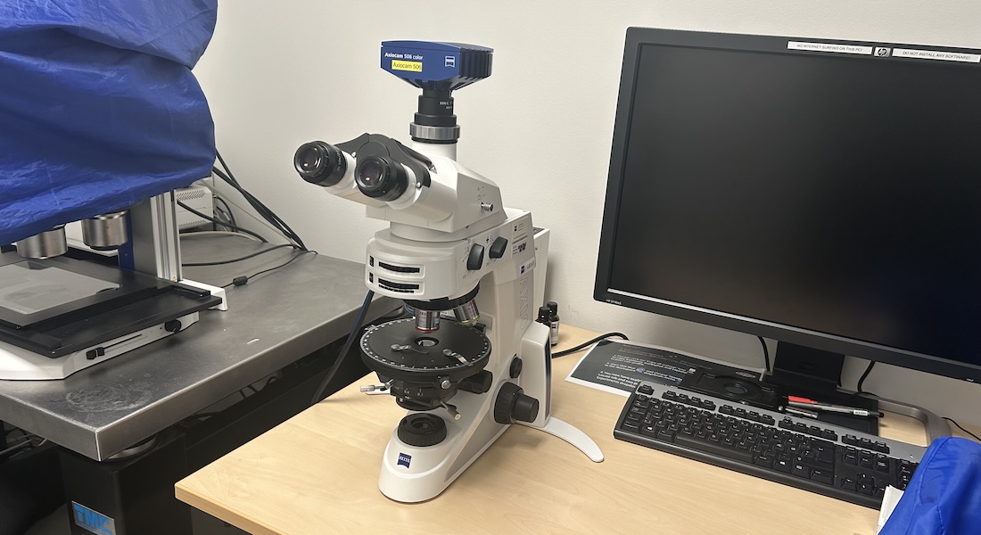
Polarization microscopy
Polarization microscopy is a contrast technique taking advantage of the optical properties of birefringent materials like bone, collagen, tendons... Birefringent objects interact differently with light depending its polarization and orientation creating contrast in the sample. Polarised light microscopy does not require any staining but works only with anisotropic samples.
Specifications
| Imaging |
Brightfield microscope equipped for polarization microscopy |
| Detectors |
AxioCam 506 6Mpx colour camera. |
| Stand |
Upright Zeiss Axiolab.A1 |
| Stage | Manual rotating stage |
| Software |
Zeiss Zen Blue 2012 |
| Illumination | Transmitted light |
| Contrast |
Brightfield Polarized light with linear or circular polarization Conoscopy |
Objectives
| Objective | Mag. / NA | Medium | Specifications | Scaling* |
| N-Achroplan | 5x / 0.15 Pol | Air | ∞ / - | 0.908µm/pix |
| N-Achroplan | 10x / 0.25 Pol | Air | ∞ / - | 0.454µm/pix |
| N-Achroplan | 20x / 0.45 Pol | Air | ∞ / 0.17 | 0.227µm/pix |
| N-Achroplan | 50x / 0.8 Pol | Air | ∞ / 0.17 | 0.0908µm/pix |
* As the nosepiece is manual, the recorded images have no scaling information. The values here were both measured from images of a stage micrometer and calculated from the hardware specifications.
Applications
Brightfield microscopy
transmitted illumination for non-fluorescent stainings such as HE, ISH...
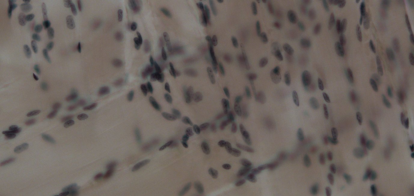
Section of a tongue. Click on the image to open in full size.
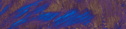
Polarization microscopy
birefringent samples interact with polarized light differently.
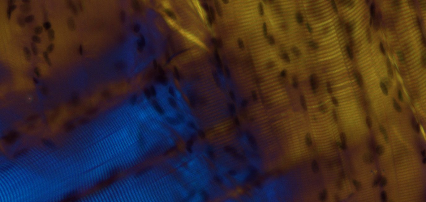
Same section as above, in polarized light microscopy. The muscles appear in different colour depending on their orientation. Click on the image to open in full size.