
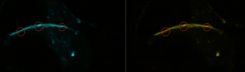
FRET by photobleaching of the acceptor. Upon photobleaching of Cx43-YFP (right), the intensity of Cx43-CFP increases (left).
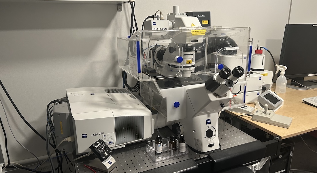
Zeiss Confocal microscope LSM 780
Laser scanning confocal microscopy has become the golden standard in imaging biological samples, allowing for good resolution and flexibility in stainings.
LSM780 is an inverted laser scanning confocal microscope equipped for live imaging (temperature and CO2 control). It has a tunable laser giving it a high flexibility in fluorophore excitation. The GaASP detector allows for detection of faint stainings and spectral imaging (separation of spectrally overlapping fluorophores).
Specifications
| Imaging | Point Scanning confocal |
| Detectors |
32-channels GaAsP detector 2 PMTs for Fluorescence (PMT2 is cooled) |
| Detection | From 405nm to 750nm |
| Stand |
INVERTED Zeiss AxioObserver Z1 |
| Stage | Motorized in XYZ |
| Software | Zeiss Zen Black 2012 |
| Add. features | Incubator (temperature + gas control) for live cell imaging |
Objectives
| Objective | Mag. / NA | Medium | Contrast | Working Distance |
| EC Plan-Neofluar | 10x / 0.3 | Air | - | 5.2 mm |
| LD Plan Neofluar | 20x / 0.8 | Air | - | Adjustable |
| LD Plan Neofluar | 40x / 0.6 | Air | - | Adjustable |
| C Apochromat | 40x / 1.2 | Water | - | Adjustable |
| EC Plan-Neofluar | 40x / 1.3 | Oil | DIC II | 0.21 mm |
| Plan-Apochromat | 63x / 1.4 | Oil | DIC III | 0.19 mm |
Light sources
| Lasers | Type and Power | Some dyes and use |
| 405nm | Diode, 30mW | DAPI, Hoechst, ... |
| 458nm | Argon, 25mW | CFP |
| 488nm | Argon, 25mW | GFP, AlexaFluor488, Cy2 |
| 514nm | Argon, 25mW | YFP |
| 543nm | HeNe, 1.2mW | RFPs, AlexaFluor568 |
| 633nm | HeNe, 5mW | DRACQ5, Cy5, AlexaFluor647 |
| Intune 488 - 645nm | 5mW |
Main beam splitters
| MBS 405/490c/640c | MBS 458 |
| MBS 405/505c | MBS 458/514 |
| MBS 405/520c | MBS 458/543 |
| MBS 405/535c | MBS 488 |
| MBS 405/550c | MBS 488/543 |
| MBS 405/565c | MBS 488/543/633 |
| MBS 405/580c | T80/R20 |
| MBS 405/595c | |
| MBS 405/610c | |
| MBS 405/625c |
Filter cubes for Mercury lamp (HXP120)
| Fluorochrome | Excitation | Dichroic | Emission | Filter No |
| Blue | SP365 | FT395 | BP420-470 | 49 |
| Green | BP470/40 | FT495 | BP525/50 | 38HE |
| Red | BP550/25 (HE) | FT570 | BP605/70 |
Applications
- Optical z-sectioning of living specimens and fixed samples,
- FRET/FRAP/FLIP/FLIM experiments,
- Photoactivation and photoconversion experiments,
- Spectral imaging and advanced unmixing of overlapping emission spectra,
- Overlay of multiple fluorescence images with transmitted light images,
- Fluorescence Correlation Spectroscopy (FCS) (up to 6 channels),
- Raster Image Correlation Spectroscopy (RICS).