
LS7 Light Sheet
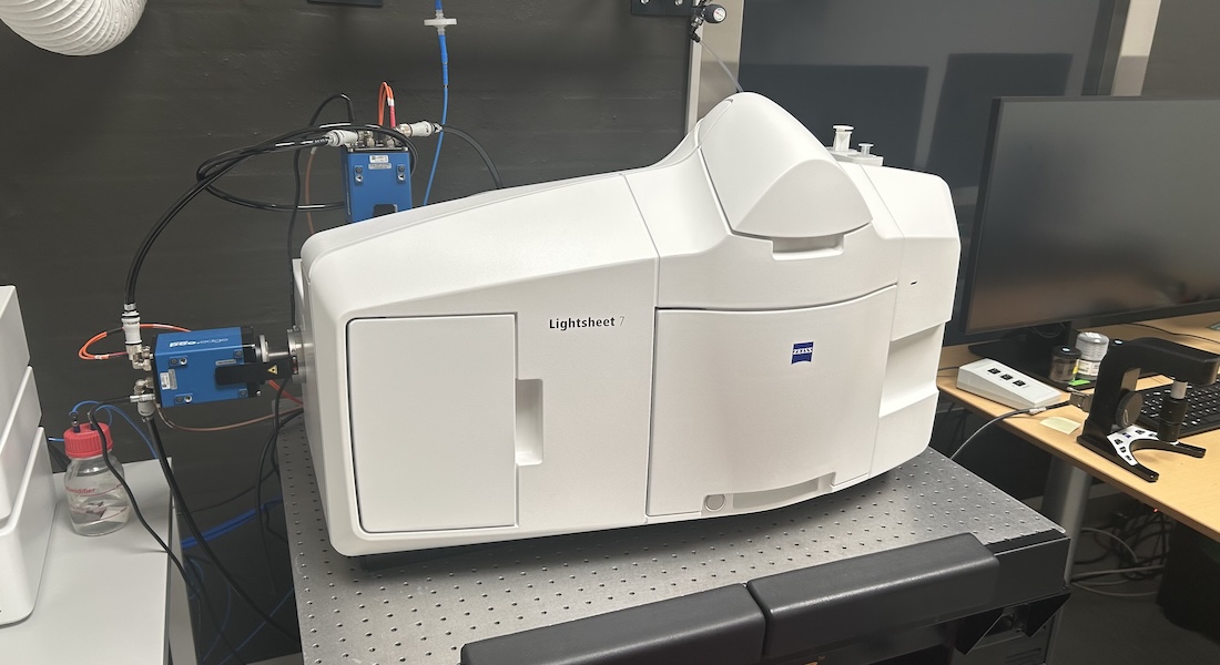 Thanks to the support from the Novo Nordisk Foundation (BioDEEP research infrastructure grant), CFIM offers open access to Light Sheet Microscopy. The Zeiss LS7 is a flexible Gaussian-beam light sheet (LS) or single plane illumination microscopy (SPIM), capable of optimally imaging small and large samples (from organoids to full embryos), both live (over time) or fixed (cleared) samples at great depths, thanks to its wide objective selection. Light sheet technologies enable for fast, gentle and deep imaging of large and/or sensitive samples while providing excellent optical sectioning due to the nature of single plane illumination.
The system boasts two illumination objectives (5x and 10x) that create a two-sided Gaussian light sheet, and a single detection objective (to choose from 5x, 10x or 20x) placed perpendicularly to the illumination plane. Signal detection is achieved by means of two Edge 4.2 sCMOS cameras by PCO. The lasers provided for excitation are 405, 488, 561 and 638 nm. This enables for collection of large sample volumes from multiple individual focal planes at various levels of detail, from mesoscopic imaging to single cell resolution.
Specifications
|
Imaging |
Gaussian-beam Light Sheet microscope |
|
Detectors |
2 PCO Edge 4.2 M sCMOS cameras |
|
Detection range |
From 405nm to 750nm |
|
Configuration |
Detection perpendicular to illumination Left and right selective plane illumination Beam pivoting to avoid shadows Objective chambers for dry and dipping lenses, and for aqueous or clearing media |
|
Software |
Zeiss Zen Black version 3.1 |
|
Add. features |
Incubation environment for live imaging Sample holders allowing multiple mounting geometries |
Objectives
|
Illumination objective |
Mag. / NA |
Medium |
Working Distance |
|
LSFM |
5x/0.1 |
Air |
N/A |
|
LSFM |
10x / 0.2 |
Air |
N/A |
|
Detection objectives |
Mag. / NA |
Medium |
Working Distance |
|
EC Plan-NEO |
5x/0.16 |
Air |
10.5 mm |
|
Plan Apo |
Water |
3.7 mm |
|
|
Plan Apo |
Water |
1.8 mm |
|
|
CIr PN |
nd=1.45 |
5.6 mm |
|
|
CIr PN |
nd=1.53 |
6.4 mm |
|
|
Cir Plan Apo |
nd=1.38 |
5.6 mm |
Light sources
|
Lasers |
Type and Power |
Some dyes and use |
|
405 nm |
Diode, 20 mW |
DAPI, Hoechst, AlexaFluor405 |
|
488 nm |
Diode, 30 mW |
GFP, AlexaFluor488, Cy2 |
|
561 nm |
Diode, 20 mW |
RFPs, AlexaFluor568, Cy3 |
|
638 nm |
Diode, 75 mW |
DRACQ5, Cy5, AlexaFluor647 |
Main beam splitter
405/488/561/638
Emission filters
|
Fluorochrome emission color |
Spectral bands |
|
|
Blue/Green |
BP 420-470 + BP 505-545 |
|
|
Green/Red |
BP 505-545 + BP 575-615 |
|
|
Green/Cherry |
BP 505-545 + LP 585 |
|
|
Green/Far Red |
BP 505-545 + LP 660 |
|
|
Red/Far Red |
BP 575-615 + LP 660 |
Applications
- Optical z-sectioning of large samples,
- 3D imaging,
- Deep imaging by using dipping lenses,
- Imaging of cleared fixed samples,
- Imaging of live small samples (organoids, embryos.