
Olympus ScanR3 Spinning Disk
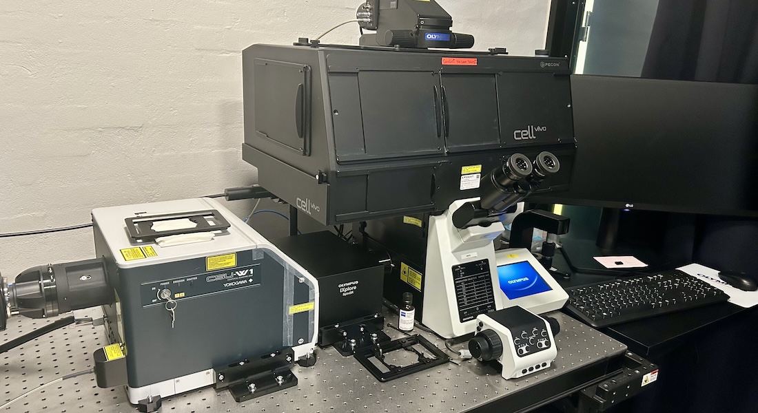
Inverted spinning disk confocal microscope designed for high-throughput content screening. Features fully automated image acquisition and data analysis. Equipped with hardware autofocusing, motorized focus and automatic XY stage.
Specifications
|
Imaging |
Spinning disk confocal and widefield microscope |
|
Detectors |
Camera (Hamamatsu Orca flash 4) Chip: Effective number of pixels: 2048x2048 Pixel Size: 6.5μm |
|
Stand |
Inverted |
|
Stage |
Motorized in XYZ |
|
Software |
ScanR Acquisition cellSens |
|
Add. features |
Temperature and CO2 Control Fully automated screening microscope Maerzhaeuser motorized stage (resolution 0.01μm, reproducibility 1.0μm, accuracy 3.0μm) Autofocus: Hardwarefocus 785nm (ZDC)/Continuous ZDC and Softwarefocus Multi Level Scan |
|
|
Yokogawa CSU-W1Confocal spinning disk unit: Microlens array 50μmdisc |
Objectives
|
Objective |
Mag. / NA |
Medium |
WD |
|
UPLXAPO 4x0.16 |
4x/0.16 |
Air |
16 |
|
UPLXAPO 10x0.4 |
10x/0.4 |
Air |
3.1 |
|
UPLXAPO 20x0.80 |
20x/0.8 |
Air |
0.6 |
|
UPLXAPO 40x0.95 |
40x/0.95 |
Air |
0.18 |
Light Sources
Lumencor Spectra LED´s
Lasers
|
Lasers |
Type and Power |
Some dyes and use |
|
405 nm |
Diode, 50 mW |
DAPI, Hoechst, AlexaFluor405, DyLight405 |
|
488 nm |
Diode, 100 mW |
GFP, AlexaFluor488, Cy2 |
|
561 nm |
Diode, 100 mW |
RFPs, AlexaFluor568, Cy3 |
|
640 nm |
Diode, 100 mW |
DRACQ5, Cy5, AlexaFluor647 |
Fluorescent filter cubes
SPX-TSEM:
- Triple band Fluorescence mirror unit
- Triple band excitation filter and dichroic beamsplitter, three single band emission filters
- Navy blue excitation (458 nm) for CFP and similar
- Emerald excitation (515 nm) for YFP and similar
- Orange excitation (594 nm) for mCherry and similar
SPX-QSEM:
- Quad band Fluorescence mirror unit
- Quad band dichroic beamsplitter, four single band emission filters
- Violet excitation (<410 nm) for DAPI and similar
- Blue excitation (480 nm) for EGFP, FITC and similar
- Yellow excitation (560 nm) for RFP and similar
- Red excitation (640 nm) for Cy 5 and similar
Applications
- Fluorescence multichannel imaging of fixed and live cells
- Fully automated image acquisition
- High throughput screening
- High content screening