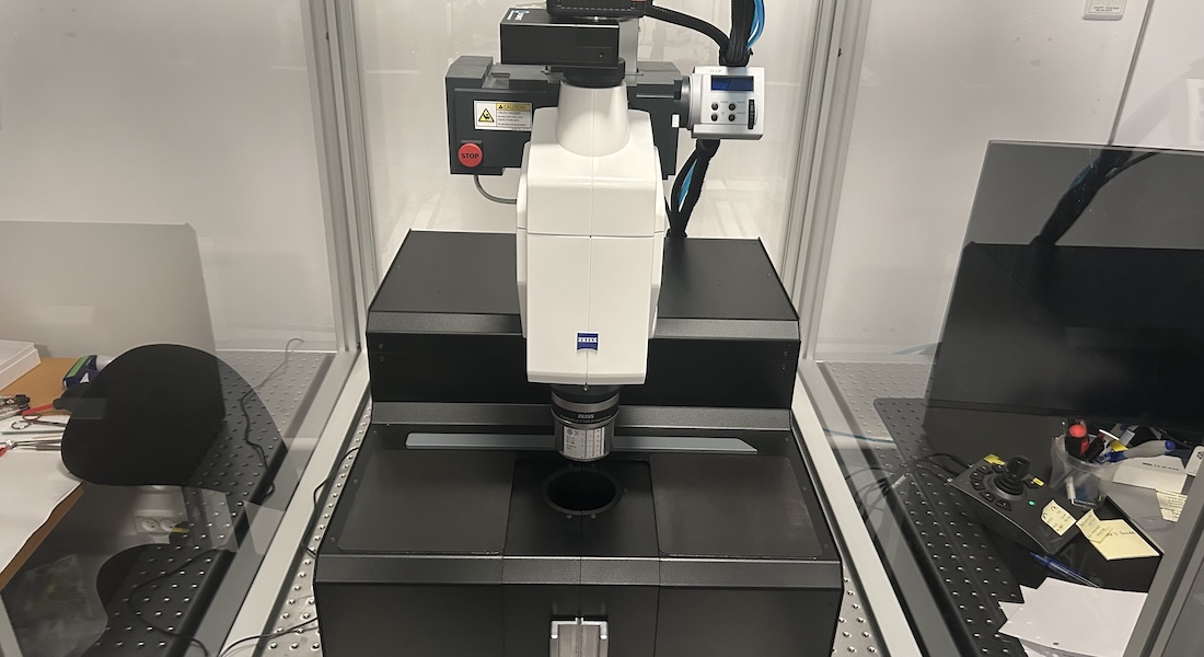
3i AxL Cleared Tissue LightSheet (CTLS)

Introduction
AxL Cleared Tissue LightSheet (CTLS) is a light sheet fluorescence microscope optimized for imaging optically cleared samples, from whole mouse organs to small whole animals. It features axially swept light sheet microscopy (ASLM) technique, scanning the thinnest part of the light sheet across large field of view in sync with the sCMOS camera rolling shutter readout, resulting in improved axial resolution.
Compatible with a range of refractive indices (RI; 1.33 - 1.58) and clearing methods, including CUBIC, iDISCO+, 2ECi, and EZ Clear.
Link to 3i AxL CTLS product information page: https://www.intelligent-imaging.com/axl
Excitation
LaserStack v4 488nm, 150mW
LaserStack v4 561nm, 150mW
LaserStack v4 637nm, 140mW
LaserStack v4 785nm, 300mW
Emission filters
446/523/600/677nm quad-band bandpass filter
525/40 nm single bandpass filter
600/52 nm single bandpass filter
692/40 nm single bandpass filter
835/70 nm single bandpass filter
Camera
Hamamatsu ORCA-Fusion BT sCMOS camera with 2304x2304 6.5µm pixels, 95% QE
Objectives
Illumination objectives:
0.14 NA illumination objectives (3i custom), spherical aberration corrected, compatible with refractive index (RI) 1.33 - 1.58.
Detection objectives:
1.0× / 0.25 NA (default)
1.5× / 0.37 NA
2.3× / 0.57 NA (with dipping cap)
Sample chambers
|
Chamber Type |
Max Sample Size (W × L × H, mm) |
Chamber Volume (mL) |
Compatible Sample Holders |
|
Small |
10 × 20 × 10 |
150 |
Small |
|
Medium |
25 × 25 × 20 |
250 |
Small, Medium |
|
Large |
25 × 50 × 20 |
500 |
Small, Medium, Large |
|
Extra Large |
30 × 100 × 30 |
1000 |
Small, Medium, Large, Extra Large |
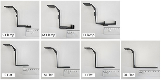
Sample holders

Tissue clearing consultation
Would like to image a large sample using light sheet microscope but new to tissue clearing? Contact us for tissue clearing consultation, customize a clearing protocol for your sample type and experiment: Sunny Dai, keqing.sunny.dai[at]sund.ku.dk
