
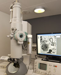
CM 100 (a & b)
Both CM 100 microscopes fulfil the purpose of standard TEM ultrastructural investigations.
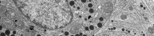
The CM100 (a) TWIN is used for conventional and negatively stained samples. The CM100 (b) has a high-contrast BioTWIN objective lens which ensures excellent contrast, even in inherently low-contrast and beam-sensitive biological specimens. Both microscopes are equipped with a side-mounted Olympus Veleta camera with a resolution of 2048 x 2048 pixels (2k x 2K).
Specifications
Filament type:
- CM100 (a): Tungsten emitter
- CM100 (b): LaB6 emitter
Accelerating Voltage (HT):
- 40KV - 100kV
Magnification range:
- CM100 (a): 18 x - 450.000 x
- CM100 (b): 18 x - 330.000 x
Applications (for both systems)
Multiple Image Alignment (MIA),
allows you to record a serie of images which are stitched together into one large image. The benefit of an MIA image over a single image recorded at a lower magnification is that you get higher resolution.
Low-Dose Technique
is used if there is a risk of damaging the specimen with the electron beam. Reducing the dose of electrons on the sample and focusing at a different spot than the area of interest are the key features to limit sample damage.
Image recording and processing
is done via the ITEM software. You need to save your images directly on your personal external HD/USB flash drive. The microscope PC is not connected to the SUND network.