New Technologies at CFIM
The Core Facility for Integrated Microscopy got new equipment, which many will hopefully benefit from. The facility has e.g. acquired new lightning-fast, super-resolution light microscopes and the ideal computer and software for 3D and 4D image data.
Watch a two minute introduction to the general facilities at CFIM.
CFIM, the faculty's joint microscopy facility, has purchased equipment that is likely to make the eyes of a lot of researchers working with imaging data shine.
Read about the new equipment below, and contact CFIM or reserve the equipment.
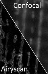
Meet the New Generation of Laser-Scanning Microscopes and configure with Airyscan 2
 CFIM has acquired two new super-resolution (above the diffraction limit) laser-scanning microscopes.
LSM980 and LSM900 represent the latest new generation of Carl Zeiss confocal microscopes, which are among the best in the world. They are ideal for fast imaging and offer high sensitivity and spectral flexibility with respect to the imaging spectrum.
To complement the microscopes, CFIM has also purchased the latest compatible configuration system, Airyscan 2. The new system allows you to obtain super-resolution imaging data of larger fields and faster than ever before. It is also equipped with a new intelligent multiplex mode, which helps you focus on the aspect of your data that is relevant to you. Read more about LSM980 Airyscan2 and LSM900 Airyscan2.
Use it for: e.g clearly resolving the intracellular compartments' boundaries.
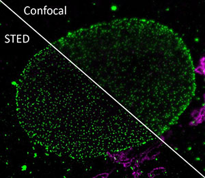
Innovatory, Compact and Very Stable STED System
 Stimulated emission depletion microscopy (STED) is a microscopy technique that creates super-resolution images through selective deactivation of fluorophores.
At CFIM, the new technique is now available in the form of the STED system called STEDYCON. STEDYCON is an innovatory, compact and very stable system that combines the STED laser and excitation laser in one beam, which makes it virtually alignment free. The system is equipped with a 775-nm STED laser and four modulated excitation laser lines (405, 488, 561, 633 nm) which, together with four APD photon-counting detectors, make it possible to produce two-coloured, super-resolution 2D images of e.g. proteins.Read more about STEDYCON.
Use it for: E.g. resolving how proteins arrange within small intracellular structures.
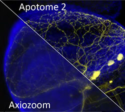
Switch Between Overview and the Smallest Details of Large Samples
 The one-axis zoom microscope, axio Zoom.V16, offers high resolution and a zoom range of 16x. It allows you to zoom seamlessly from overview to the smallest details and to combine images fast.
Moreover, the Apotome.2 module enables optical sectioning of large samples. This structured illumination technique enables imaging of thicker samples without excessive out-of-focus blur. Read more at cfim.ku.dk.
Use it for: E.g. imaging of large fluorescent samples in 3D.
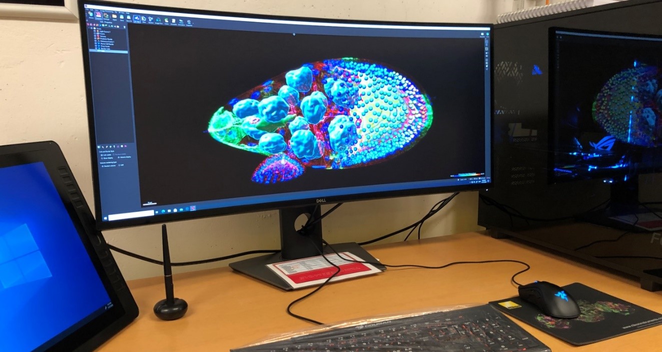
Superfast Work Station with the Ideal Software
Meet Ramon y Cajal – our new supercomputer for processing imaging data. It is optimal for large data sets and highly demanding image processing and visualisation tasks. The work station is equipped with IMARIS for Cell Biologists software, which is the best tool in the world for studying and analysing living cells and for processing 3D and 4D data. It is also equipped with Huygens Professional, which is ideal for deconvolution of light microscopy images.
Specifications: 64-core AMD Threadripper 3990x, 256-GB RAM, 14-TB storage and GeForce 2080Ti graphics card.
Use it for: Image processing and analysis of big data sets.

