Lightsheet Imaging Workshop

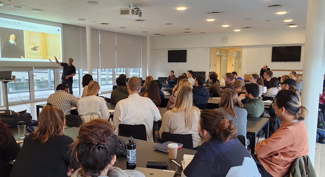
Exploring the three-dimensional organization of intact tissues has a high impact in the biomedical research field. Lightsheet fluorescence microscopy (LSFM) has arisen as a powerful method for efficient 3D imaging of live and fixed specimens. Its high acquisition speed and low bleaching, combined with the new tissue clearing techniques, enables imaging from millimeters to centimeters of tissue, revealing the full 3D structure of the specimen without sectioning.
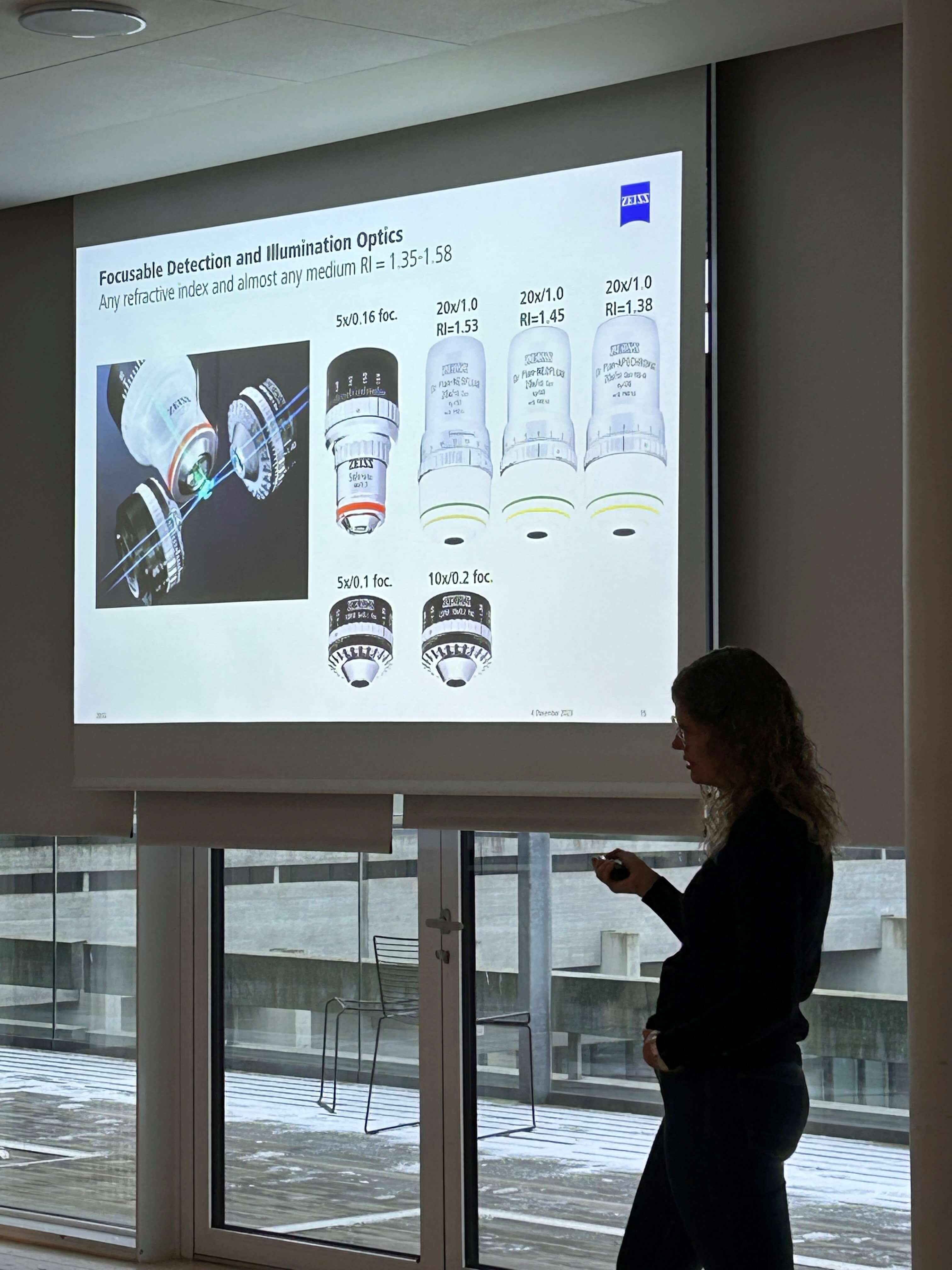
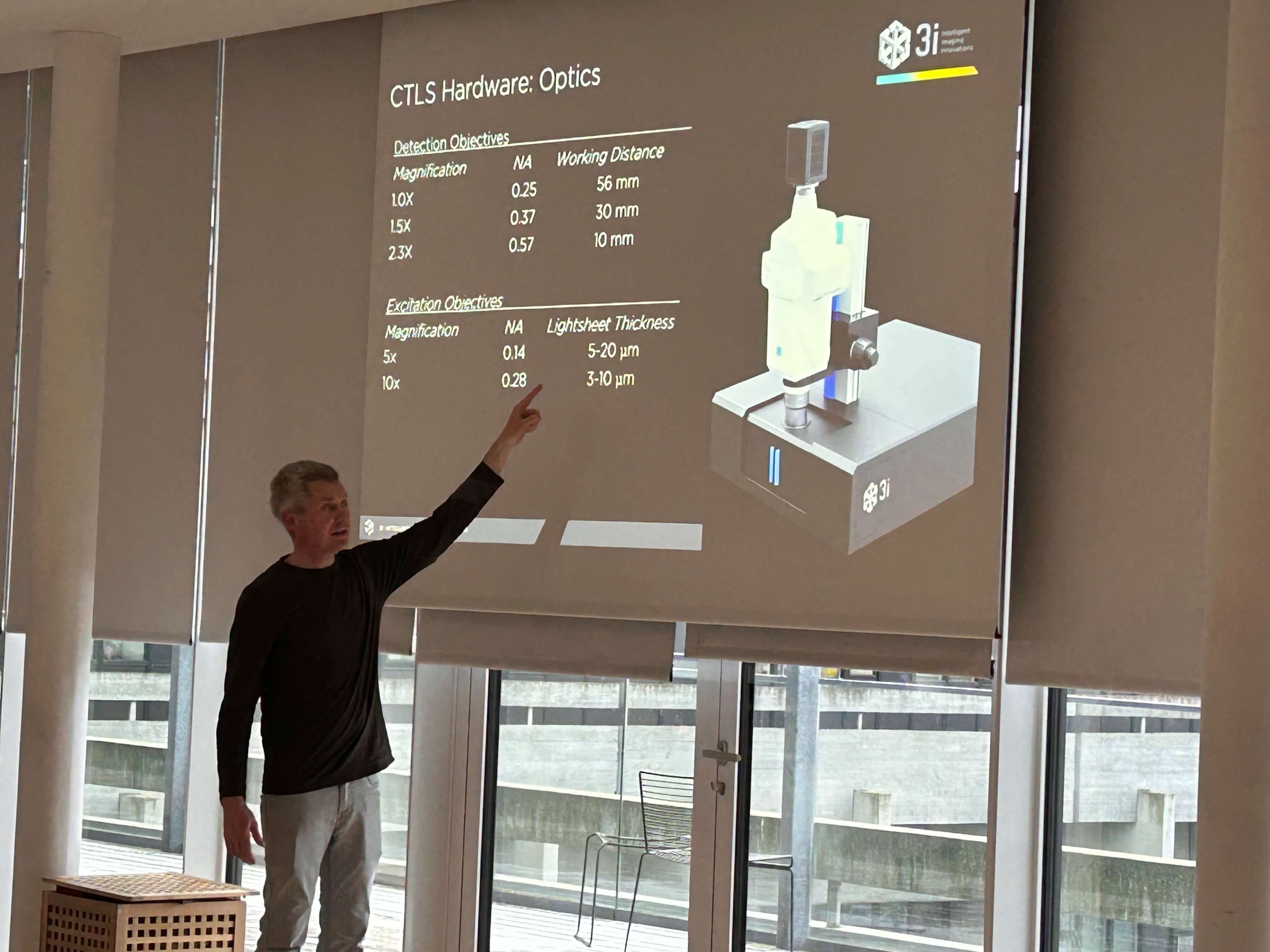
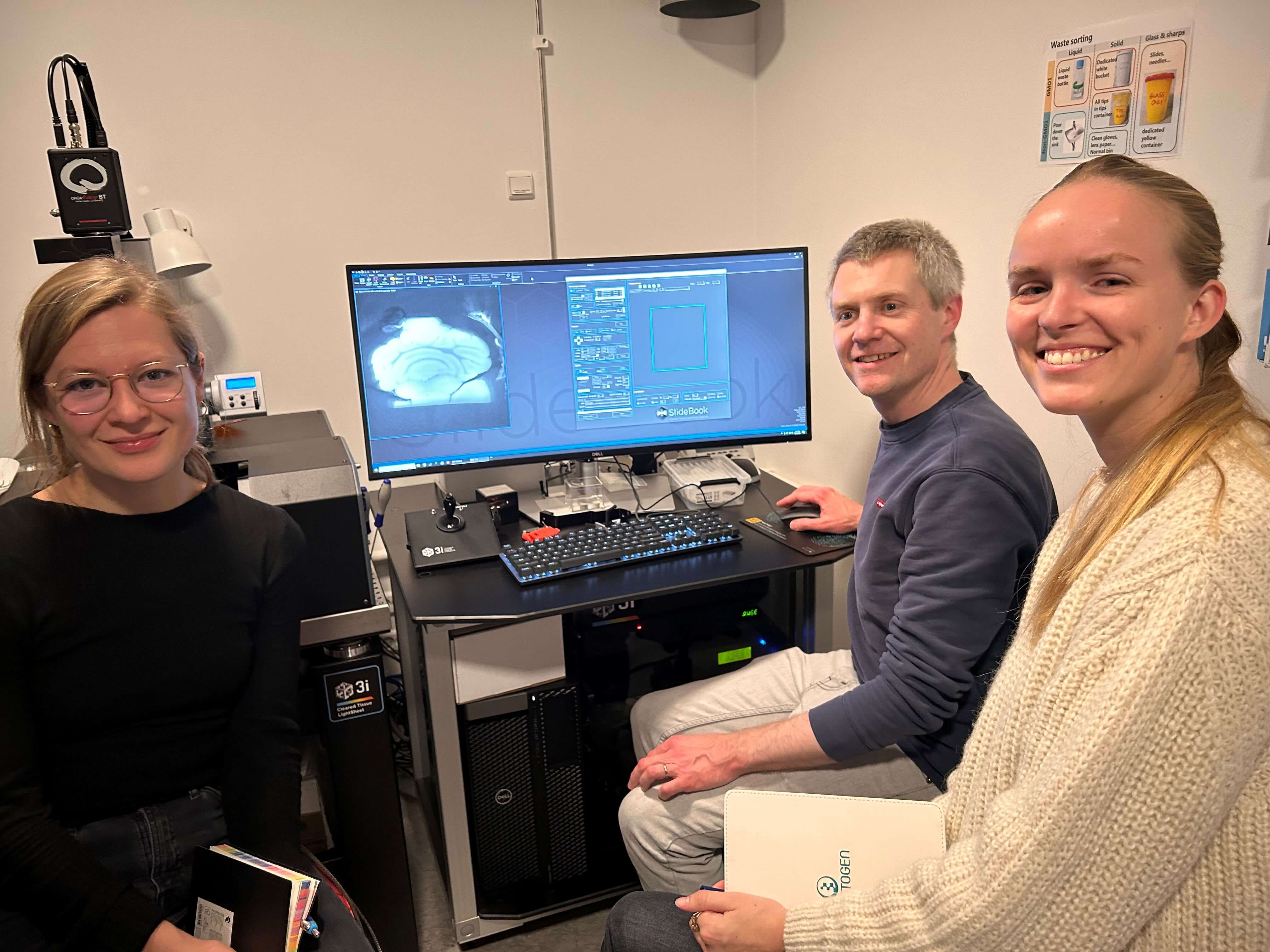
In this workshop, we will focus on the advantages and the main challenges in the state-of-the-art clearing techniques. We will demonstrate the LSFM imaging capabilities with readymade cleared samples, using the 3i Cleared Tissue LightSheet and the Zeiss LS7 microscopes.
In addition, we will briefly show different approaches for the large data handling, analysis and rendering.
The workshop will start with lectures open to 80 participants, followed by hands on sessions for 16 participants on clearing methods, and image acquisition
with both microscopes.
If you are interested, you could discuss the experimental design of your project with the leading experts from Zeiss and 3i and bring your own samples for imaging.


