
EM sample preparation lab
CFIM's sample preparation lab is equipped with many high-end instruments. These cannot be booked individually but are used when a specimen preparation is ordered (under booking > service).
Tissue Processor for Electron and Light Microscopy Resin Processing
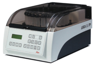
The Leica EM TP is a tissue processor designed for EM and LM resin processing that features a heating/cooling system with pre-heat and pre-cool capabilities to maintain a constant processing temperature.
It includes an easy-to-operate membrane control panel and the sample carousel holds 24 EM or 12 LM vials and utilizes various specialized baskets and capsules. An exhaust system supports safer use of toxic substances.
Automatic Contrasting Instrument for Electron Microscopy
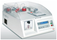
Photo credit: Leica
The Leica EM AC20 automatic contrasting system for ultra-thin sections ensures minimum user contact with reagents and reduced reagent consumption.
Usage of a peristaltic pump and non-contact valves allow the reagents to travel directly through tubing to the grid chamber, resulting in fast, high-quality double staining and easy maintenance.
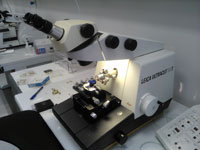
Ultra microtome for Precise Room Temperature and Cryo Sectioning
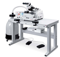
Photo credit: Leica
The Leica Ultra microtome UC7 provides easy preparation of semi- and ultra-thin sections as well as perfect, smooth surfaces of biological samples for TEM, SEM, AFM and LM examination.
With Cryo chamber FC7 mounted to the UC7 Ultra microtome, we are also able to make cryo-sections (-15° to -185°C) for TEM, SEM, AFM and LM examination.
Knife Making
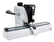
Photo credit: Leica
Leica EM KMR3 makes glass knives for perfect ultrathin sections for EM and LM applications.
The balanced break method of the EM KMR3 ensures perfect glass knives in three thicknesses; 6.4 mm, 8 mm and 10 mm.
The EM KMR3 is easy to use combined with the automatic reset of the breaking wheel and scoring mechanism to "default" after a breaking cycle, avoids handling errors.
Ultramicrotomes
 Leica Ultracut UCT and Reichert Ultracut S ultramicrotomes are instruments commonly used to prepare TEM samples. They use either a glass or diamond knife to cut ultra thin sections of specimens embedded in epoxy. The sections are approximately 60 to 100 nanometers in thickness and 0.5 to 3 mm in width and can be stained and mounted onto TEM grids for viewing.