
Tecnai G2 20 TWIN

The FEI Tecnai G2 20 TWIN is a 200 kV Transmission Electron Microscope.
It has an advanced operating system: all microscope components - the electron gun, the optical elements, the vacuum system and the stage - are digitally controlled by the Tecnai user interface on the microscope PC. Data acquisition is done with the bottom mounted FEI High-Sensitive (HS) 4k x 4k Eagle camera or the Side-mounted Olympus 1.3k x 1K Megaview G2 camera. Both camera’s are embedded in the Tecnai user interface and can be automated for acquisition processes, like Cryo with low-dose technique & tomography.
The Tecnai is also equipped with the iCorr which is a fully integrated wide-field fluorescence microscope. iCorr allows to image sequentially with fluorescence and electron microscopy, without manually changing the sample between the 2 modes (Correlative Light and Electron Microscopy: an Impression and the Solutions).
Specifications
- Filament type: LaB6 emitter
- Accelerating Voltage (HT): 40KV - 200kV
- Magnification range: 25 x - 700.000 x
Applications
Correlative microscopy
is done in combination with the iCorr. By tilting the sample holder 90° degrees, you can fast localize precise areas of interest using the fluorescence microscope and then tilt the holder back to 0° degrees and look for ultra-structural details with the electron microscope.
The fluorescence signal in an image identifies the positions where labeled molecules or structures of interest are located. These positions can be stored with a click of a mouse using the iCorr software. Using a shared coordinate system, these positions of interest are automatically located and imaged in TEM mode to gather ultra-structural detail with a single mouse click. These TEM images are automatically superimposed on the LM grid overview with precise accuracy, delivering fast, high quality correlated results.
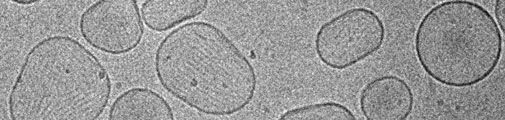
Cryo-applications
are used to examine frozen suspensions of single particles, 2D protein crystal sheets and frozen-hydrated sections.
The microscope is equipped with retractable liquid-nitrogen cooled Gatan Cryo-blades and it shields the specimen almost completely off from residual gases so that the ice growth rate has been significant reduced on cold specimens inside the microscope. For on-site cryo sample preparation, a FEI Vitrobot tm Mark IV is used, which has a fully PC-controlled touch screen and is used for vitrification (rapid cooling) of aqueous samples in liquid ethane to eliminate ice crystal formation during sample vitrification.
Low-Dose Technique
is used if there is a risk of damaging the specimen, such as biological samples preserved either in vitreous ice or epoxy resin, with the electron beam. Reducing the dose of electrons on the sample and focusing at a different spot than the area of interest are the key features to limit sample damage.
Tomography
is used to obtaining detailed 3D structures of sub cellular macromolecular objects. The electron beam is passing through the sample at incremental degrees of rotation (max ± 65 degrees) around the center of the target sample (tilt axis). The acquired tilt series is used to assemble a three dimensional image of the target and this is done with the Xplore3D software package.
This application consists of three modules; one for the acquisition of tomography data on the Tecnai (tilt series acquisition), one for alignment and reconstruction of the acquired tilt series (Inspect3D), the last one is to visualize the reconstructed 3D image (Amira), rapid exploration and analysis of 3D data and generating polygonal and tetrahedral 3D models for advanced visualization and simulation.
Another option for reconstruction of your acquired tilt series are the following software packages: IMOD and FiJi (ImageJ in combination with java3D and special plug-ins). The image post processing software (except for IMOD) is featured on all PCs in the CFIM meeting room (21.01.24A).