
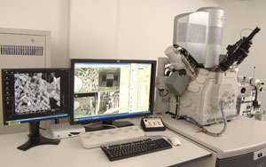
Quanta FEG 3D
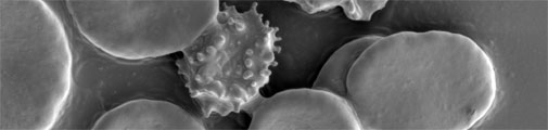
The FEI Quanta 3D FEG is a versatile high-resolution dualbeam SEM for 2D & 3D characterization and analysis. The microscope combines innovative electron and ion optics with Quanta's environmental mode (ESEM). Optimized for high brightness and high beam current operation, this ultra-stable electron field emission gun (FEG) significantly extends the use of high spatial resolution.
The high-current FIB enables fast material removal and automated FIB sectioning programs enable accurate cross-sectioning.
With the arrival of the new MAPS software, the gap between light and electron microscopy has never been as narrow as ever before. Impression and solutions for Correlative Light and Electron Microscopy (CLEM).
Specifications
- E-beam Imaging features a pre-aligned electron optical column which is optimized for high resolution and beam stability.
- The Quanta has a resolution in high-vacuum mode with the E-beam of 1.2 nm at 30kV with the SED detector
- Accelerating voltages E-beam: 200V up to 30kV.
- Magnification range: 30 x up to 1.280.000 x
- Vacuum levels: Through-the-lens differential pumping apertures resulting 3 different vacuum pressure levels; High-Vacuum, Low-Vacuum and ESEM (environmental). The low and ESEM-vacuum capability enables almost charge free imaging and analysis of non-conductive specimens and/or hydrated specimens.
- Specimen stage: A high-precision, 5 axes motorized specimen stage with 50 mm travel along the X and Y axes, with a maximal sample height of 50 mm at an optimum tilt of 52 degrees for FIB technique.
- FIB, is a high current Focused Ion Beam. The FIB acts as a ‘nano scalpel’ enabling high precision cutting & slicing (up to 10 nm/slice) into samples to reveal their 3D internal structure. The Quanta is equipped with a gas injection system (GIS) for Platinum (PT) deposition on to the sample.
- Cryo applications: the system is equipped with the Leica Cryo stage and uses the EM VCT100 Cryo transport system, in combination with the MED020 freeze fracture and Platinum/Carbon or Carbon coating unit.
- TEM sample Prep integrated Omni-probe for TEM sample lift-out.
- Operating system, windows XP based pc with a 4-quadrant User Interface, digital image processing up to 4k x 4k image resolution.
Detectors
- SED, Secondary Electrons are detected with the following detectors.
- High vacuum mode: Everhart-Thornley Detector(ETD)
- ESEM (gaseous) mode : Low-vacuum Detector (Lv-SED) and Gaseous detector (GSED) - vCD, retractable 4-segment backscattered electron detector (BSED) which operate at low voltage and with high contrast.
- iCD, In-column backscattered electron detector (BSED).
- IR camera, Integrated Infra-Red inspection camera for viewing at the sample/column inside the chamber
Software
- Auto Slice and View software for unattended serial sectioning and imaging through a site-specific volume of the specimen. The sequence of images can be merged into a video or be used as the input for 3-dimensional reconstruction.
- 3D reconstruction the Amira software package allows rapid exploration and analysis of 3D data, as well as for generation of polygonal and tetrahedral 3D models for advanced visualization and simulation.
- Correlative microscopy is done with the CorrSight and the MAPS software. Import and correlate image data from any external imaging source (i.e., light microscope) through an easy-to-use interface, resulting in data visibility ranging from low- magnification surface imaging (LM) to high-magnification, extreme resolution images at nanoscale (EM).